
Understanding ECG
What is an ECG ?
ECG, or electrocardiogram, is the graphical representation of the electrical activity of the heart. It can detect certain cardiovascular pathologies.
With each heartbeat, an electrical wave travels through your heart. This wave causes your heart to contract and pump blood.
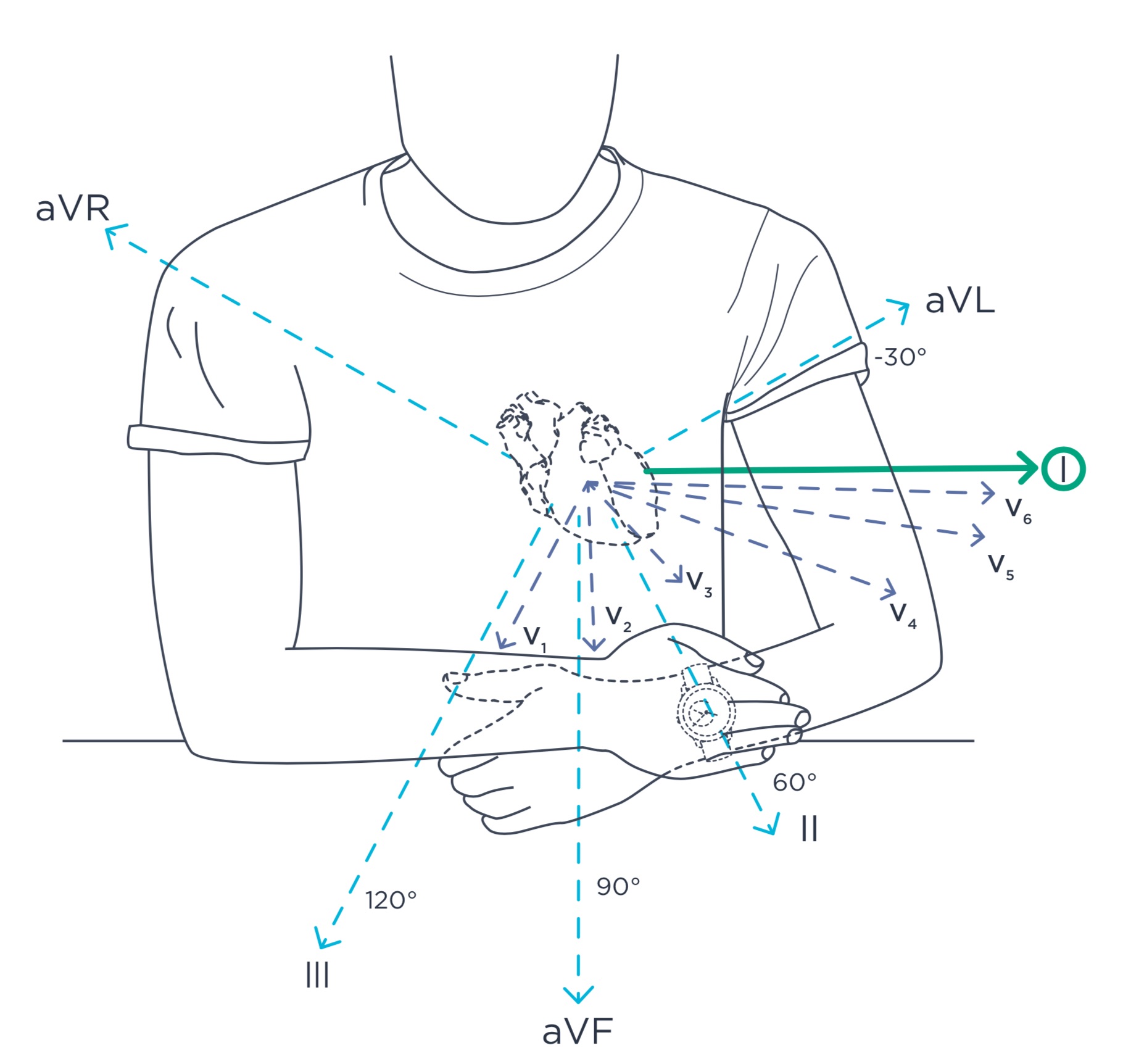
The gold standard ECG records 12 lead (or ways) of the heart. Withings ECG devices sense the electrical activity of the heart at a distance through the left and right arm. This configuration is known as lead D1.
What practitioners look for on the ECG graph
A normal heartbeat on the ECG graph will show the time the electric wave takes to move through your heart. There are 3 distinct patterns:
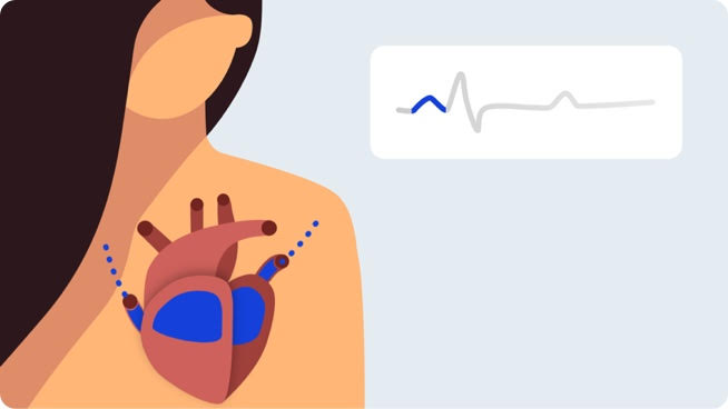
- First Wave: The "P wave"
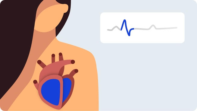
- Next Wave: the "QRS Complex"
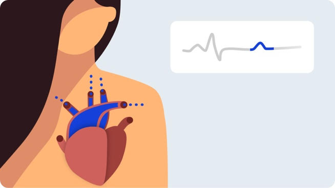
- Final Wave: the "T wave"
The graph shows a first electric wave as electricity moves into the right and left upper chambers of your heart.
This second pattern shows electricity moving through the right and left bottom chambers.
The final wave represents electrical recovery or return to a resting state for the ventricles.
By measuring time intervals on an ECG, practitioners can find out if this electricity travel is too slow, normal, too fast, or even irregular.
Which conditions can Withings ECG devices detect ?
ECGs can detect a range of conditions, from angina pectoris to severe heart attacks. Withings ECG devices focus on detecting atrial fibrillation. If you believe you are having a heart attack, contact emergency services.
Results you may see
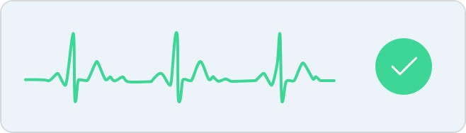
Normal Sinus Rhythm
A sinus rhythm means your heart is beating in a uniform pattern.
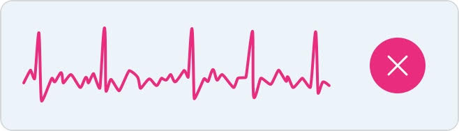
Atrial fibrillation
Atrial fibrillation occurs when the two upper chambers of the heart move chaotically instead of pumping regularly. The P wave on the ECG disappears and is replaced by a jumpy baseline. The QRS complex occurs at "irregularly irregular" intervals.
Inconclusive
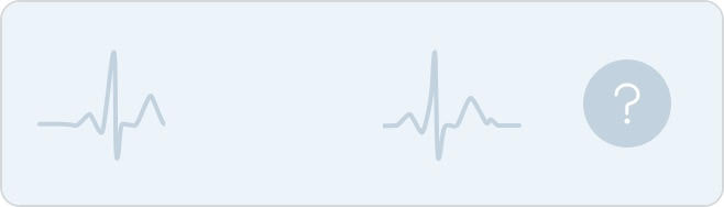
An inconclusive result means the recording can’t be classified. This can happen for many reasons.
Heart rate is low: the heart rate obtained cannot be classified as a recording. To obtain a full analysis, the heart rate must be above 50 bpm during the recording.
Heart rate is high: a complete diagnosis is not possible for a heart rate above 100 bpm. To obtain a full analysis, the heart rate must be below 100 bpm during the recording.
Signs of other arrhythmia: an inconclusive signal can be explained by the presence of an arrhythmia other than atrial fibrillation, or a undle branch block.
Noise
Signal is too noisy: there is too much interference for the recording to be classified. Place your arm on a table or on your thigh, relax, don’t talk, and don’t move during the recording. Refer to the best practices section to know the right gestures to be adopted and those to be avoided.
If you think you may be having a heart attack (myocardial infarction) or are facing a medical emergency, call the emergency services.